What Provides the Information Necessary to Specify the Three-dimensional Shape of a Protein?

Protein structure is the three-dimensional organization of atoms in an amino acid-concatenation molecule. Proteins are polymers – specifically polypeptides – formed from sequences of amino acids, the monomers of the polymer. A unmarried amino acid monomer may also be called a residual indicating a repeating unit of a polymer. Proteins grade by amino acids undergoing condensation reactions, in which the amino acids lose one water molecule per reaction in gild to attach to one some other with a peptide bond. Past convention, a chain under thirty amino acids is often identified every bit a peptide, rather than a protein.[ane] To be able to perform their biological function, proteins fold into i or more specific spatial conformations driven by a number of non-covalent interactions such as hydrogen bonding, ionic interactions, Van der Waals forces, and hydrophobic packing. To sympathise the functions of proteins at a molecular level, it is often necessary to decide their three-dimensional structure. This is the topic of the scientific field of structural biology, which employs techniques such equally X-ray crystallography, NMR spectroscopy, cryo electron microscopy (cryo-EM) and dual polarisation interferometry to determine the construction of proteins.
Protein structures range in size from tens to several yard amino acids.[2] By physical size, proteins are classified as nanoparticles, betwixt i–100 nm. Very large protein complexes tin be formed from protein subunits. For example, many thousands of actin molecules assemble into a microfilament.
A protein unremarkably undergoes reversible structural changes in performing its biological function. The alternative structures of the same protein are referred to every bit different conformations, and transitions between them are chosen conformational changes.
Levels of protein structure [edit]
There are iv distinct levels of protein structure.
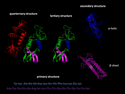
Four levels of poly peptide structure
Master structure [edit]
The main structure of a protein refers to the sequence of amino acids in the polypeptide chain. The primary structure is held together past peptide bonds that are made during the procedure of protein biosynthesis. The two ends of the polypeptide chain are referred to as the carboxyl terminus (C-terminus) and the amino terminus (Due north-terminus) based on the nature of the free grouping on each extremity. Counting of residues ever starts at the North-terminal stop (NHii-group), which is the cease where the amino group is non involved in a peptide bail. The primary structure of a protein is adamant by the factor corresponding to the protein. A specific sequence of nucleotides in Deoxyribonucleic acid is transcribed into mRNA, which is read past the ribosome in a process called translation. The sequence of amino acids in insulin was discovered by Frederick Sanger, establishing that proteins have defining amino acid sequences.[3] [4] The sequence of a protein is unique to that protein, and defines the structure and function of the poly peptide. The sequence of a protein can be adamant by methods such as Edman degradation or tandem mass spectrometry. Often, notwithstanding, it is read direct from the sequence of the cistron using the genetic code. It is strictly recommended to utilize the words "amino acid residues" when discussing proteins considering when a peptide bond is formed, a water molecule is lost, and therefore proteins are made up of amino acid residues. Post-translational modifications such as phosphorylations and glycosylations are usually also considered a part of the master structure, and cannot be read from the gene. For case, insulin is composed of 51 amino acids in two chains. One concatenation has 31 amino acids, and the other has 20 amino acids.
Secondary structure [edit]
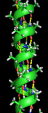
An α-helix with hydrogen bonds (yellow dots)
Secondary construction refers to highly regular local sub-structures on the actual polypeptide courage chain. Two chief types of secondary structure, the α-helix and the β-strand or β-sheets, were suggested in 1951 by Linus Pauling et al.[5] These secondary structures are divers past patterns of hydrogen bonds betwixt the main-chain peptide groups. They have a regular geometry, existence constrained to specific values of the dihedral angles ψ and φ on the Ramachandran plot. Both the α-helix and the β-sheet represent a way of saturating all the hydrogen bond donors and acceptors in the peptide backbone. Some parts of the protein are ordered only exercise not form any regular structures. They should non be dislocated with random coil, an unfolded polypeptide chain lacking whatsoever fixed 3-dimensional structure. Several sequential secondary structures may form a "supersecondary unit".[6]
Third structure [edit]
Tertiary structure refers to the three-dimensional construction created by a single protein molecule (a single polypeptide chain). It may include one or several domains. The α-helixes and β-pleated-sheets are folded into a compact globular construction. The folding is driven by the non-specific hydrophobic interactions, the burying of hydrophobic residues from water, merely the structure is stable only when the parts of a protein domain are locked into place by specific tertiary interactions, such as salt bridges, hydrogen bonds, and the tight packing of side chains and disulfide bonds. The disulfide bonds are extremely rare in cytosolic proteins, since the cytosol (intracellular fluid) is generally a reducing surround.
Quaternary structure [edit]
Quaternary construction is the 3-dimensional construction consisting of the aggregation of ii or more individual polypeptide chains (subunits) that operate as a single functional unit (multimer). The resulting multimer is stabilized by the same non-covalent interactions and disulfide bonds as in 3rd construction. There are many possible 4th structure organisations.[vii] Complexes of ii or more polypeptides (i.e. multiple subunits) are called multimers. Specifically it would be called a dimer if it contains two subunits, a trimer if information technology contains three subunits, a tetramer if it contains four subunits, and a pentamer if it contains five subunits. The subunits are oft related to one another by symmetry operations, such as a two-fold axis in a dimer. Multimers made upward of identical subunits are referred to with a prefix of "homo-" and those made upward of different subunits are referred to with a prefix of "hetero-", for example, a heterotetramer, such equally the two alpha and ii beta bondage of hemoglobin.
Domains, motifs, and folds in protein structure [edit]

Proteins are frequently described as consisting of several structural units. These units include domains, motifs, and folds. Despite the fact that there are about 100,000 different proteins expressed in eukaryotic systems, there are many fewer different domains, structural motifs and folds.
Structural domain [edit]
A structural domain is an chemical element of the poly peptide'southward overall structure that is self-stabilizing and oftentimes folds independently of the residuum of the poly peptide chain. Many domains are not unique to the poly peptide products of one cistron or one gene family but instead appear in a diverseness of proteins. Domains oft are named and singled out considering they effigy prominently in the biological function of the protein they belong to; for case, the "calcium-binding domain of calmodulin". Because they are independently stable, domains can be "swapped" past genetic engineering science between one protein and another to make chimera proteins. A conservative combination of several domains that occur in dissimilar proteins, such equally protein tyrosine phosphatase domain and C2 domain pair, was called "a superdomain" that may evolve every bit a single unit.[eight]
Structural and sequence motifs [edit]
The structural and sequence motifs refer to short segments of protein iii-dimensional structure or amino acid sequence that were found in a big number of different proteins
Supersecondary structure [edit]
The supersecondary construction refers to a specific combination of secondary construction elements, such equally β-α-β units or a helix-turn-helix motif. Some of them may be also referred to equally structural motifs.
Protein fold [edit]
A poly peptide fold refers to the general protein architecture, like a helix bundle, β-barrel, Rossmann fold or unlike "folds" provided in the Structural Nomenclature of Proteins database.[9] A related concept is protein topology.
Protein dynamics and conformational ensembles [edit]
Proteins are not static objects, but rather populate ensembles of conformational states. Transitions between these states typically occur on nanoscales, and have been linked to functionally relevant phenomena such as allosteric signaling[x] and enzyme catalysis.[11] Protein dynamics and conformational changes permit proteins to function equally nanoscale biological machines inside cells, oft in the form of multi-protein complexes.[12] Examples include motor proteins, such as myosin, which is responsible for muscle contraction, kinesin, which moves cargo inside cells away from the nucleus along microtubules, and dynein, which moves cargo inside cells towards the nucleus and produces the axonemal chirapsia of motile cilia and flagella. "[I]northward result, the [motile cilium] is a nanomachine composed of perhaps over 600 proteins in molecular complexes, many of which too office independently as nanomachines...Flexible linkers let the mobile protein domains continued past them to recruit their binding partners and induce long-range allostery via protein domain dynamics. "[thirteen]
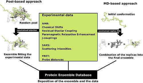
Schematic view of the two main ensemble modeling approaches.[14]
Proteins are oft thought of as relatively stable tertiary structures that experience conformational changes later on being afflicted by interactions with other proteins or equally a role of enzymatic activity. Still, proteins may have varying degrees of stability, and some of the less stable variants are intrinsically disordered proteins. These proteins exist and function in a relatively 'disordered' state lacking a stable tertiary structure. As a result, they are difficult to describe by a single stock-still third structure. Conformational ensembles accept been devised as a mode to provide a more than accurate and 'dynamic' representation of the conformational country of intrinsically disordered proteins.[15] [xiv]
Poly peptide ensemble files are a representation of a protein that can exist considered to have a flexible structure. Creating these files requires determining which of the various theoretically possible protein conformations actually be. I approach is to apply computational algorithms to the protein data in order to try to determine the about probable set of conformations for an ensemble file. There are multiple methods for preparing data for the Protein Ensemble Database that fall into two general methodologies – pool and molecular dynamics (MD) approaches (diagrammed in the figure). The pool based approach uses the protein's amino acid sequence to create a massive pool of random conformations. This pool is then subjected to more computational processing that creates a ready of theoretical parameters for each conformation based on the construction. Conformational subsets from this pool whose average theoretical parameters closely match known experimental data for this protein are selected. The alternative molecular dynamics approach takes multiple random conformations at a fourth dimension and subjects all of them to experimental information. Hither the experimental data is serving as limitations to be placed on the conformations (e.k. known distances between atoms). Only conformations that manage to remain within the limits set past the experimental data are accepted. This approach often applies large amounts of experimental data to the conformations which is a very computationally demanding chore.[14]
The conformational ensembles were generated for a number of highly dynamic and partially unfolded proteins, such as Sic1/Cdc4,[16] p15 PAF,[17] MKK7,[18] Beta-synuclein[nineteen] and P27[20]
Protein folding [edit]
| | This section needs expansion. You tin can help by adding to information technology. (Apr 2019) |
As it is translated, polypeptides exit the ribosome mostly every bit a random coil and folds into its native land.[21] [22] The final structure of the poly peptide concatenation is generally assumed to exist determined by its amino acrid sequence (Anfinsen's dogma).[23]
Protein stability [edit]
Thermodynamic stability of proteins represents the free free energy departure between the folded and unfolded protein states. This gratis energy difference is very sensitive to temperature, hence a change in temperature may result in unfolding or denaturation. Protein denaturation may result in loss of function, and loss of native country. The free free energy of stabilization of soluble globular proteins typically does not exceed 50 kJ/mol.[ citation needed ] Taking into consideration the big number of hydrogen bonds that have place for the stabilization of secondary structures, and the stabilization of the inner core through hydrophobic interactions, the complimentary free energy of stabilization emerges as small divergence between large numbers.[24]
Protein structure determination [edit]

Examples of protein structures from the PDB
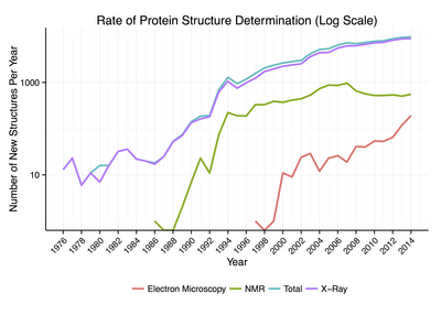
Rate of Protein Construction Determination past Method and Year
Around 90% of the protein structures bachelor in the Protein Information Banking concern have been adamant by X-ray crystallography.[25] This method allows one to measure the three-dimensional (3-D) density distribution of electrons in the protein, in the crystallized state, and thereby infer the iii-D coordinates of all the atoms to be determined to a certain resolution. Roughly nine% of the known protein structures have been obtained by nuclear magnetic resonance (NMR) techniques.[ commendation needed ] For larger protein complexes, cryo-electron microscopy can determine poly peptide structures. The resolution is typically lower than that of Ten-ray crystallography, or NMR, just the maximum resolution is steadily increasing. This technique is still a peculiarly valuable for very large protein complexes such as virus coat proteins and amyloid fibers.
General secondary construction composition tin can be determined via circular dichroism. Vibrational spectroscopy can also exist used to characterize the conformation of peptides, polypeptides, and proteins.[26] Two-dimensional infrared spectroscopy has get a valuable method to investigate the structures of flexible peptides and proteins that cannot be studied with other methods.[27] [28] A more qualitative film of protein structure is often obtained by proteolysis, which is also useful to screen for more crystallizable protein samples. Novel implementations of this arroyo, including fast parallel proteolysis (FASTpp), can probe the structured fraction and its stability without the demand for purification.[29] Once a protein's structure has been experimentally determined, further detailed studies can be washed computationally, using molecular dynamic simulations of that structure.[xxx]
Protein structure databases [edit]
A protein structure database is a database that is modeled around the diverse experimentally determined protein structures. The aim of nigh protein structure databases is to organize and comment the protein structures, providing the biological community access to the experimental information in a useful fashion. Data included in protein structure databases ofttimes includes 3D coordinates as well as experimental information, such equally unit cell dimensions and angles for x-ray crystallography adamant structures. Though well-nigh instances, in this case either proteins or a specific structure determinations of a poly peptide, as well contain sequence information and some databases even provide means for performing sequence based queries, the primary attribute of a structure database is structural data, whereas sequence databases focus on sequence data, and contain no structural information for the majority of entries. Protein structure databases are critical for many efforts in computational biology such every bit structure based drug design, both in developing the computational methods used and in providing a large experimental dataset used past some methods to provide insights about the part of a protein.[31]
Structural classifications of proteins [edit]
Poly peptide structures tin be grouped based on their structural similarity, topological class or a common evolutionary origin. The Structural Classification of Proteins database[32] and CATH database[33] provide two different structural classifications of proteins. When the structural similarity is big the two proteins accept possibly diverged from a mutual ancestor,[34] and shared structure between proteins is considered evidence of homology. Structure similarity can then be used to grouping proteins together into protein superfamilies.[35] If shared structure is significant but the fraction shared is modest, the fragment shared may be the event of a more than dramatic evolutionary outcome such every bit horizontal gene transfer, and joining proteins sharing these fragments into protein superfamilies is no longer justified.[34] Topology of a protein can be used to classify proteins also. Knot theory and excursion topology are 2 topology frameworks developed for classification of protein folds based on chain crossing and intrachain contacts respectively.
Computational prediction of protein construction [edit]
The generation of a protein sequence is much easier than the determination of a poly peptide structure. However, the structure of a protein gives much more insight in the function of the protein than its sequence. Therefore, a number of methods for the computational prediction of poly peptide structure from its sequence accept been adult.[36] Ab initio prediction methods use just the sequence of the protein. Threading and homology modeling methods can build a 3-D model for a poly peptide of unknown structure from experimental structures of evolutionarily-related proteins, called a poly peptide family.
See also [edit]
- Biomolecular structure
- Gene structure
- Nucleic acrid structure
- PCRPi-DB
- Ribbon diagram 3D schematic representation of proteins
References [edit]
- ^ H. Stephen Stoker (1 Jan 2015). Organic and Biological Chemistry. Cengage Learning. p. 371. ISBN978-1-305-68645-8.
- ^ Brocchieri L, Karlin Due south (10 June 2005). "Protein length in eukaryotic and prokaryotic proteomes". Nucleic Acids Research. 33 (10): 3390–3400. doi:10.1093/nar/gki615. PMC1150220. PMID 15951512.
- ^ Sanger, F.; Tuppy, H. (one September 1951). "The amino-acid sequence in the phenylalanyl chain of insulin. I. The identification of lower peptides from partial hydrolysates". The Biochemical Periodical. 49 (4): 463–481. doi:10.1042/bj0490463. ISSN 0264-6021. PMC1197535. PMID 14886310.
- ^ Sanger, F. (fifteen May 1959). "Chemical science of Insulin". Science. 129 (3359): 1340–1344. Bibcode:1959Sci...129.1340G. doi:x.1126/science.129.3359.1340. ISSN 0036-8075. PMID 13658959.
- ^ Pauling L, Corey RB, Branson Hr (1951). "The structure of proteins; two hydrogen-bonded helical configurations of the polypeptide concatenation". Proc Natl Acad Sci USA. 37 (4): 205–211. Bibcode:1951PNAS...37..205P. doi:x.1073/pnas.37.four.205. PMC1063337. PMID 14816373.
- ^ Chiang YS, Gelfand TI, Kister AE, Gelfand IM (2007). "New classification of supersecondary structures of sandwich-similar proteins uncovers strict patterns of strand aggregation". Proteins. 68 (4): 915–921. doi:10.1002/prot.21473. PMID 17557333. S2CID 29904865.
- ^ Moutevelis Eastward, Woolfson DN (January 2009). "A periodic table of coiled-whorl protein structures". J. Mol. Biol. 385 (3): 726–32. doi:ten.1016/j.jmb.2008.11.028. ISSN 0022-2836. PMID 19059267.
- ^ Haynie DT, Xue B (2015). "Superdomain in the protein construction hierarchy: the case of PTP-C2". Protein Science. 24 (5): 874–82. doi:ten.1002/pro.2664. PMC4420535. PMID 25694109.
- ^ Govindarajan Southward, Recabarren R, Goldstein RA (17 September 1999). "Estimating the total number of protein folds". Proteins. 35 (iv): 408–414. doi:ten.1002/(SICI)1097-0134(19990601)35:4<408::AID-PROT4>3.0.CO;2-A. hdl:2027.42/34969. PMID 10382668. Archived from the original on 5 Jan 2013.
- ^ Bu Z, Callaway DJ (2011). "Proteins Motion! Protein dynamics and long-range allostery in cell signaling". Protein Structure and Diseases. Advances in Poly peptide Chemistry and Structural Biology. Vol. 83. Academic Press. pp. 163–221. doi:x.1016/B978-0-12-381262-9.00005-7. ISBN9780123812629. PMID 21570668.
- ^ Fraser JS, Clarkson MW, Degnan SC, Erion R, Kern D, Alber T (December 2009). "Hidden culling structures of proline isomerase essential for catalysis". Nature. 462 (7273): 669–673. Bibcode:2009Natur.462..669F. doi:10.1038/nature08615. PMC2805857. PMID 19956261.
- ^ Donald, Voet (2011). Biochemistry. Voet, Judith Thousand. (fourth ed.). Hoboken, NJ: John Wiley & Sons. ISBN9780470570951. OCLC 690489261.
- ^ Satir, Peter; Søren T. Christensen (26 March 2008). "Structure and part of mammalian cilia". Histochemistry and Cell Biological science. 129 (6): 687–93. doi:10.1007/s00418-008-0416-nine. PMC2386530. PMID 18365235. 1432-119X.
- ^ a b c Varadi, Mihaly; Vranken, Wim; Guharoy, Mainak; Tompa, Peter (one January 2015). "Computational approaches for inferring the functions of intrinsically matted proteins". Frontiers in Molecular Biosciences. 2: 45. doi:10.3389/fmolb.2015.00045. PMC4525029. PMID 26301226.
- ^ Protein Ensemble Database
- ^ Mittag, Tanja; Marsh, Joseph; Grishaev, Alexander; Orlicky, Stephen; Lin, Hong; Sicheri, Frank; Tyers, Mike; Forman-Kay, Julie D. (14 March 2010). "Structure/part implications in a dynamic complex of the intrinsically matted Sic1 with the Cdc4 subunit of an SCF ubiquitin ligase". Structure. xviii (4): 494–506. doi:10.1016/j.str.2010.01.020. ISSN 1878-4186. PMC2924144. PMID 20399186.
- ^ De Biasio, Alfredo; Ibáñez de Opakua, Alain; Cordeiro, Tiago N.; Villate, Maider; Merino, Nekane; Sibille, Nathalie; Lelli, Moreno; Diercks, Tammo; Bernadó, Pau (18 Feb 2014). "p15PAF is an intrinsically disordered protein with nonrandom structural preferences at sites of interaction with other proteins". Biophysical Journal. 106 (4): 865–874. Bibcode:2014BpJ...106..865D. doi:10.1016/j.bpj.2013.12.046. ISSN 1542-0086. PMC3944474. PMID 24559989.
- ^ Kragelj, Jaka; Palencia, Andrés; Nanao, Max H.; Maurin, Damien; Bouvignies, Guillaume; Blackledge, Martin; Jensen, Malene Ringkjøbing (17 March 2015). "Structure and dynamics of the MKK7-JNK signaling complex". Proceedings of the National Academy of Sciences of the U.s. of America. 112 (11): 3409–3414. Bibcode:2015PNAS..112.3409K. doi:ten.1073/pnas.1419528112. ISSN 1091-6490. PMC4371970. PMID 25737554.
- ^ Allison, Jane R.; Rivers, Robert C.; Christodoulou, John C.; Vendruscolo, Michele; Dobson, Christopher G. (25 November 2014). "A human relationship betwixt the transient structure in the monomeric state and the aggregation propensities of α-synuclein and β-synuclein". Biochemistry. 53 (46): 7170–7183. doi:10.1021/bi5009326. ISSN 1520-4995. PMC4245978. PMID 25389903.
- ^ Sivakolundu, Sivashankar G.; Bashford, Donald; Kriwacki, Richard West. (11 Nov 2005). "Disordered p27Kip1 exhibits intrinsic structure resembling the Cdk2/cyclin A-bound conformation". Journal of Molecular Biology. 353 (5): 1118–1128. doi:10.1016/j.jmb.2005.08.074. ISSN 0022-2836. PMID 16214166.
- ^ Zhang, Gong; Ignatova, Zoya (1 Feb 2011). "Folding at the nascence of the nascent concatenation: coordinating translation with co-translational folding". Electric current Opinion in Structural Biology. 21 (1): 25–31. doi:x.1016/j.sbi.2010.10.008. ISSN 0959-440X. PMID 21111607.
- ^ Alberts, Bruce; Alexander Johnson; Julian Lewis; Martin Raff; Keith Roberts; Peter Walters (2002). "The Shape and Structure of Proteins". Molecular Biology of the Cell; Quaternary Edition. New York and London: Garland Scientific discipline. ISBN978-0-8153-3218-3.
- ^ Anfinsen, C. (1972). "The germination and stabilization of poly peptide construction". Biochem. J. 128 (4): 737–49. doi:ten.1042/bj1280737. PMC1173893. PMID 4565129.
- ^ Jaenicke, R.; Heber, U.; Franks, F.; Chapman, D.; Griffin, Mary C. A.; Hvidt, A.; Cowan, D. A. (1990). "Poly peptide Structure and Function at Low Temperatures [and Discussion]". Philosophical Transactions of the Royal Society of London. Serial B, Biological Sciences. 326 (1237): 535–553. doi:10.1098/rstb.1990.0030. JSTOR 2398703. PMID 1969647.
- ^ Kendrew, J.C.; Bodo, M.; Dintzis, H. Thou.; Parrish, R. Grand.; Wyckoff, H.; Phillips, D.C. (1958). "A Three-Dimensional Model of the Myoglobin Molecule Obtained by 10-Ray Assay". Nature. 181 (4610): 662–666. Bibcode:1958Natur.181..662K. doi:10.1038/181662a0. PMID 13517261. S2CID 4162786.
- ^ Krimm Southward, Bandekar J (1986). "Vibrational spectroscopy and conformation of peptides, polypeptides, and proteins". Advances in Protein Chemistry Volume 38. Adv. Protein Chem. Advances in Protein Chemistry. Vol. 38. pp. 181–364. doi:10.1016/S0065-3233(08)60528-8. ISBN9780120342389. PMID 3541539.
- ^ Lessing, J.; Roy, S.; Reppert, M.; Baer, Chiliad.; Marx, D.; Jansen, T.L.C.; Knoester, J.; Tokmakoff, A. (2012). "Identifying Residue Structure in Intrinsically Matted Systems: A second IR Spectroscopic Study of the GVGXPGVG Peptide". J. Am. Chem. Soc. 134 (11): 5032–5035. doi:ten.1021/ja2114135. hdl:11370/ff19c09b-088a-48f0-afee-2111a9b19252. PMID 22356513.
- ^ Jansen, T.Fifty.C.; Knoester, J. (2008). "2-dimensional infrared population transfer spectroscopy for enhancing structural markers of proteins". Biophys. J. 94 (five): 1818–1825. Bibcode:2008BpJ....94.1818J. doi:10.1529/biophysj.107.118851. PMC2242754. PMID 17981904.
- ^ Minde DP, Maurice MM, Rüdiger SG (2012). "Determining biophysical protein stability in lysates by a fast proteolysis assay, FASTpp". PLOS ONE. vii (x): e46147. Bibcode:2012PLoSO...746147M. doi:x.1371/journal.pone.0046147. PMC3463568. PMID 23056252.
- ^ Kumari I, Sandhu P, Ahmed Chiliad, Akhter Y (August 2017). "Molecular Dynamics Simulations, Challenges and Opportunities: A Biologist'southward Prospective". Curr. Poly peptide Pept. Sci. xviii (11): 1163–1179. doi:ten.2174/1389203718666170622074741. PMID 28637405.
- ^ Laskowski, RA (2011). "Poly peptide structure databases". Mol Biotechnol. 48 (ii): 183–98. doi:10.1007/s12033-010-9372-four. PMID 21225378. S2CID 45184564.
- ^ Murzin, A. G.; Brenner, S.; Hubbard, T.; Chothia, C. (1995). "SCOP: A structural nomenclature of proteins database for the investigation of sequences and structures" (PDF). Journal of Molecular Biology. 247 (4): 536–540. doi:10.1016/S0022-2836(05)80134-2. PMID 7723011. Archived from the original (PDF) on 26 April 2012.
- ^ Orengo, C. A.; Michie, A. D.; Jones, S.; Jones, D. T.; Swindells, M. B.; Thornton, J. M. (1997). "CATH--a hierarchic classification of protein domain structures". Construction. 5 (viii): 1093–1108. doi:10.1016/S0969-2126(97)00260-viii. PMID 9309224.
- ^ a b Pascual-García, A.; Abia, D.; Ortiz, A.R.; Bastolla, U. (2009). "Cross-over between detached and continuous protein structure space: insights into automatic nomenclature and networks of protein structures". PLOS Computational Biology. five (3): e1000331. Bibcode:2009PLSCB...5E0331P. doi:ten.1371/journal.pcbi.1000331. PMC2654728. PMID 19325884.
- ^ Holm, 50; Rosenström, P (July 2010). "Dali server: conservation mapping in 3D". Nucleic Acids Research. 38 (Spider web Server issue): W545–ix. doi:10.1093/nar/gkq366. PMC2896194. PMID 20457744.
- ^ Zhang Y (2008). "Progress and challenges in poly peptide construction prediction". Curr Opin Struct Biol. 18 (3): 342–348. doi:10.1016/j.sbi.2008.02.004. PMC2680823. PMID 18436442.
Further reading [edit]
- 50 Years of Protein Construction Determination Timeline - HTML Version - National Institute of General Medical Sciences at NIH
External links [edit]
-
 Media related to Protein structures at Wikimedia Eatables
mcclellandtakether.blogspot.com
Source: https://en.wikipedia.org/wiki/Protein_structure
0 Response to "What Provides the Information Necessary to Specify the Three-dimensional Shape of a Protein?"
Post a Comment